

Password Reset
Forgot your password? Enter the email address you used to create your account to initiate a password reset.


Forgot your password? Enter the email address you used to create your account to initiate a password reset.
A study regarding operable models was recently published in the open access journal of the American Academy of Otolaryngology – Head and Neck Surgery Foundation. The study was authored by several experts, including Noel Jabbour, MD, from the UPMC Department of Otolaryngology and Anish Ghodadra, MD, from the UPMC Department of Radiology.
Deconstructing surgeries into steps and providing instructions with illustrations has been the staple of surgical textbooks for decades. However, it may be difficult for the novice surgeon to interpret 2-dimensional (2D) illustrations into 3D surgeries.
The purpose of the study is to create operable models that demonstrate the progression of surgery in 3D and allow for mastering the final steps of the operation first. Mastoidectomy was performed in a stepwise fashion to different end points on five identical 3D-printed temporal bone models to represent five major steps of the operation. The drilled models were computed tomography scanned and the subsequent images were used to create 3D model copies of each step.
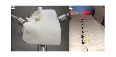
This is the first study to demonstrate that it is possible to create, scan, and copy stepwise, operable, patient-specific 3D-printed models, which the trainee can both reference as a 3D dissection guide and operate on repeatedly and in any order.
Read more about this study on OTO Open.
Collaborators not affiliated with the University of Pittsburgh:
Monika E. Freiser, MD
West Virginia University
Michael Magnetta, MD
NorthShore University Health System
Jonathan E. Castaño, MD
West Virginia University
