

Password Reset
Forgot your password? Enter the email address you used to create your account to initiate a password reset.


Forgot your password? Enter the email address you used to create your account to initiate a password reset.
9 Minutes
 The first artificial iris implant at UPMC was performed in December 2025 at the UPMC Vision Institute by Ian Conner, MD, PhD, division chief of the Vision Institute’s Glaucoma and Cataract Service and associate professor of ophthalmology at the University of Pittsburgh School of Medicine.


The recipient was Evan Facher, PhD, MBA, vice chancellor for innovation and entrepreneurship at the University of Pittsburgh and associate dean for commercial translation at the University of Pittsburgh School of Medicine, who had sustained a sports-related open-globe injury to his left eye 18 months earlier. Boris Rosin, MD, PhD, vitreoretinal surgeon at the Vision Institute and assistant professor of ophthalmology at the University of Pittsburgh School of Medicine, managed Dr. Facher’s retinal care from the primary repair through the final reconstruction.
“The artificial iris is the only option that approaches normal function for patients with large iris defects. Until now, patients in western Pennsylvania who needed one were referred elsewhere. Offering it at the UPMC Vision Institute keeps our patient’s care close to home and provides referring physicians in the region a local option for this complex procedure,” Dr. Conner says.
 Case Description
Dr. Facher presented in June 2024 after a sports-related injury to the left eye. Computed tomography showed rupture of the globe without orbital fracture. At surgical exploration, vitreous and approximately 30% of the iris had extruded through the rupture. The crystalline lens was disrupted, with lens material lost through the wound, the capsular bag and zonules were destroyed, and the retina was detaching.
“Rupture of the globe requires substantial force and often expels iris, lens, and vitreous through the wound. Tissue lost that way is contaminated and cannot be restored, so the primary repair is directed at closing the globe and preserving what remains for subsequent reconstruction,” Dr. Conner says.
 The primary repair was performed the day after the injury. Extruded tissue was excised and the globe was closed. Dr. Rosin performed pars plana vitrectomy with laser retinopexy and gas tamponade and placed a scleral buckle. Four weeks after the primary repair, Dr. Rosin removed residual lens material, performed a vitreous washout, and applied supplemental laser retinopexy.
“Traumatic traction on the peripheral retina leaves these eyes at high risk for tears and detachment while they heal. Standard management is emergency closure of the globe followed by a second operation to secure the retina. Reconstruction is considered only after the globe and retina have stabilized,” Dr. Conner says.
The globe and retina were then allowed to heal. During the interval between the initial surgeries and reconstruction, Dr. Facher was aphakic, with a large iris defect and a functionally fixed, dilated pupil, and had severe photophobia, glare, and impaired depth perception. Contact lens correction was not feasible in the traumatized eye.
“Without a functioning iris, the eye is effectively under full pharmacologic dilation at all times. Patients in this situation often keep the affected eye closed or patched to get through the day, which makes ordinary activities difficult,” Dr. Conner says.
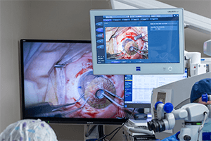 On preoperative evaluation prior to reconstruction and implantation of the artificial iris, the retina was attached and pinhole acuity indicated good visual potential. With no capsular or zonular support, Dr. Conner planned scleral fixation of an intraocular lens (IOL) and artificial iris implant in a single procedure, with Dr. Rosin performing concurrent vitreoretinal surgery.
“When the crystalline lens is present, the artificial iris can be placed in the capsular bag with the IOL during cataract surgery. In a pseudophakic eye, sulcus placement is possible, but after trauma, a passively fixated implant is unlikely to remain stable, and a second operation becomes a real possibility. Because Dr. Facher was aphakic, the IOL required scleral fixation, and suturing the iris to the sclera as well provided the most secure fixation available. We do not expect either implant to dislocate,” Dr. Conner says.
Dr. Conner sutured the IOL and the artificial iris to the sclera, passing sutures through each implant and the eye wall, and removed residual sutures from the primary repair. Dr. Rosin removed scar tissue from the posterior segment, identified a new small retinal tear, and treated it with laser retinopexy and gas tamponade.
Postoperative inflammation and surface irritation from absorbable sutures resolved over approximately two months. At four and a half months, best-corrected visual acuity in the left eye was 20/25 with spectacle correction, and photophobia had largely resolved. Dr. Facher’s follow-up care alternates between Dr. Conner and Dr. Rosin every two months, and eventually that will change to twice a year as he continues to recover from the surgery.
 “Nothing in Dr. Facher’s preoperative evaluation suggested anything other than our being able to achieve an excellent clinical outcome. The principal risks came from the operation itself, because prolonged inflammation and elevated intraocular pressure occur at a predictable rate after a third procedure on a complicated eye, and he has avoided both to date. The eye will require close surveillance for life, and I expect to see him at least every six months even if his course remains uneventful,” Dr. Conner says.
The CustomFlex® Artificial Iris is manufactured in Germany by HumanOptics AG. It was approved by the U.S. Food and Drug Administration in 2018 and remains the only device of its kind approved for use in the United States. The CustomFlex artificial iris is indicated for treating congenital aniridia and for iris defects resulting from trauma, albinism, or surgical excision. The device has no moving parts and no optic, so it is paired with a separate IOL when one is needed. The device is inserted into the eye folded through a small incision and flattened within the eye. The device’s central 3.5 mm wide aperture is the same on every device.
HumanOptics makes each implant by hand for the individual patient, with the anterior surface colored to match the individual’s fellow iris from high-resolution photographs. Manufacture and delivery of the custom device for patients takes several months.
“The implant is thin, light, and made of a silicone with established biocompatibility. Those properties are important in eyes that have already undergone trauma and multiple operations. The device has proven to be well tolerated by patients,” Dr. Conner says.
Because the aperture of the artificial iris is fixed, its size determines how the eye performs in different lighting conditions and how well the posterior segment can be examined afterward. Eyes with traumatic iris loss as happened in this case to Dr. Facher typically need retinal surveillance for life because of the increased risks for tears or detachments, and the iris aperture has to account for this long-term clinical need.
“A smaller aperture would improve glare control at the cost of vision in low light, and a larger one would do the reverse. The aperture also has to permit a thorough view of the retina because these eyes need posterior segment examination for the rest of the patient’s life. The 3.5 mm opening balances all three requirements,” Dr. Conner says.
Candidates for an artificial iris are patients with large iris defects, most often after trauma, whose eyes can otherwise support a good visual outcome. Many patients are aphakic or have lost capsular support and require scleral fixation of both an IOL and the iris implant, frequently in eyes that have already undergone vitreoretinal surgery. Device-specific certification requires documented experience in complex anterior segment surgery, didactic training, and model-eye practice, which Dr. Conner completed before bringing the procedure to UPMC.
“Artificial iris implantation is outside the scope of a routine cataract surgery practice. It depends on experience with scleral-fixated lenses and other complex anterior segment procedures that most ophthalmologists do not perform,” Dr. Conner says. “Candidates reach this procedure only through referral. Many patients who could benefit are likely still being managed in general ophthalmology practices without having been evaluated for it,” Dr. Conner says.
Health care coverage for the artificial iris right now is limited, and, therefore, it is essential that referring physicians and their patients understand the potential costs before evaluation proceeds. The implant itself costs approximately $10,000 and the device manufacturer requires payment up front before producing and shipping the device. In almost all cases, the patient must pay for the device themselves while other aspects of their surgical care may be covered by their health insurance coverage.
“I discuss the cost of the implant early in counseling, before planning is underway. For some patients, $10,000 is manageable, but for many it is a significant barrier. Patients and referring physicians should be aware of the financial obligations from the outset,” Dr. Conner says.
Dr. Facher’s case and the reconstructive surgical plan for such a complex injury depended on the extensive vitreoretinal and complex anterior segment surgery capabilities available at the UPMC Vision Institute, and a plan devised and executed by a multidisciplinary team across three operations over 18 months. The continuity of care along Dr. Facher’s entire journey highlights how the UPMC Vision Institute is structured and how various ophthalmic disciplines routinely come together to tackle complex eye conditions and injuries.
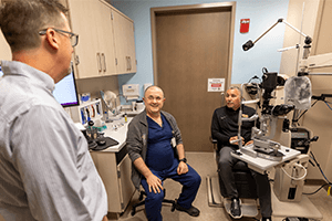 “In complex trauma, the anterior segment and the retina cannot be managed in isolation. Planning and performing the reconstruction with Dr. Rosin, who had monitored and cared for Dr. Facher’s retina since the primary repair, is what allowed for the subsequent success we have seen with the anterior segment reconstruction and artificial iris,” Dr. Conner says.
Dr. Facher, whose role at the University of Pittsburgh focuses on moving research into clinical practice, has become an advocate for the technology and for broader access to it.
“Dr. Facher’s professional background gives him an unusual understanding of what it takes to bring a technology like the artificial iris to patients. He has advocated for himself and for the device, and he has given generously of his time to help make it more widely known and available. Greater awareness benefits every patient who could be a candidate,” Dr. Conner says.
To refer a patient to Dr. Conner for artificial iris consultation, contact the UPMC Vision Institute at 412-647-2200.

