


Password Reset
Forgot your password? Enter the email address you used to create your account to initiate a password reset.


Forgot your password? Enter the email address you used to create your account to initiate a password reset.
15 Minutes
 In mid-2026, the Gamma Knife® stereotactic radiosurgery program, in the UPMC Center for Image-Guided Neurosurgery (CIGNS), treated its 20,000th patient since L. Dade Lunsford, MD, brought the first Gamma Knife in North America to UPMC and the University of Pittsburgh in 1987. In the four decades since Dr. Lunsford and colleagues treated their first patient with the novel device, the program and its voluminous experience have helped define how stereotactic radiosurgery (SRS) can be used across brain and skull-base tumors, vascular malformations, and functional disorders, while generating valuable long-term clinical data needed to determine whether those treatments and outcomes remain durable and safe decades after treatment. Today, that same legacy is supporting a new generation of research in artificial intelligence, computational neurosurgery, and individualized radiosurgical treatment planning for patients.
The Gamma Knife program at UPMC grew from Dr. Lunsford’s work in image-guided and stereotactic neurosurgery, and his advanced fellowship training in stereotactic neurosurgery with pioneering Swedish neurosurgeon Lars Leksell, MD, who invented the Gamma Knife. Dr. Lunsford, now the Lars Leksell Distinguished Professor of Neurological Surgery and director emeritus of the Center for Image-Guided Neurosurgery, remains active in research, clinician training, and advocacy.
 Bringing the Gamma Knife to Pittsburgh in the mid-1980s was no small task, conceptually, logistically, and fiscally. * While it was the first device of its kind in North America, it was only the fifth in the world. The work to bring the device to Pittsburgh included many individual partners and collaborators, university and hospital leadership, and a core team that included John C. Flickinger, MD, FACR, professor of radiation oncology and neurological surgery, and the medical physics team to develop an approach in which neurological surgery, radiation oncology, and medical physics functioned together in treatment planning and delivery.
That multidisciplinary structure, developed at UPMC, remains fundamental to how Gamma Knife treatments are planned, conducted, studied, and taught globally. Neurosurgeons contribute disease-specific and anatomical expertise; radiation oncologists contribute expertise in radiation biology, dose, and treatment effects; and medical physicists are integral to dosimetry and treatment planning. The disciplines work together rather than sequentially around one goal: devise the most optimal and safest approach to treating the individual patient in front of them.

 Constantinos G. Hadjipanayis, MD, PhD, the L. Dade Lunsford Professor, executive vice chair, co-director of the UPMC Brain Tumor Center, and director of the Brain Tumor Nanotechnology Laboratory, now directs the UPMC Center for Image-Guided Neurosurgery and helps lead the Gamma Knife program. Dr. Hadjipanayis trained at UPMC under Dr. Lunsford from 1998 through 2006, when stereotactic radiosurgery was becoming established for use in a growing number of neurological conditions. Co-directing the UPMC CIGNS is Ajay Niranjan, MD, MBA, professor of neurosurgery, director of the UPMC Brain Mapping Center, and director of radiosurgery research in the CIGNS.
The significance of the program’s nearly 40-year history and treating the 20,000th patient means so much more than just a procedural number tally. It’s not the number itself that matters. It’s what that number translates to in terms of clinical knowledge and the evolution of the clinical use of Gamma Knife radiosurgery, and research questions that can be asked and answered – questions that only time and large clinical volumes can inform. When the Gamma Knife arrived in Pittsburgh, it was not possible to know for certain whether radiosurgery could control a meningioma or vestibular schwannoma for decades, if it could replace surgery for complex intracranial lesions, or if adverse effects would emerge 10, 20, or 30 years after treatment that would render its use obsolete. Answering those kinds of questions requires sustained clinical experience, institutional continuity, and long-term follow-up. What began as a pioneering treatment has become one of the most extensively studied approaches in modern neurosurgery. Decades of follow-up have allowed UPMC researchers to evaluate long-term tumor control, neurological outcomes, treatment-related complications, and the durability of radiosurgical interventions. These findings have continuously refined patient selection and treatment strategies, transforming clinical experience into scientific evidence that continues to shape contemporary radiosurgery.
“When this technology came here in 1987, the idea that you could treat an intracranial lesion and send the patient home the same day seemed almost futuristic. I would characterize it as Dr. Lunsford, the hospital, and university supporting the introduction of Gamma Knife as an informed leap of faith,” Dr. Hadjipanayis says. “Dr. Lunsford had a visionary outlook on the world of neurosurgery with image-guided neurosurgery and stereotactic radiosurgery that others did not have at the time. There was no way to know what these patients would look like 20 or 30 years later. Now we have the follow-up to know what happens to these tumors and other conditions over decades after Gamma Knife treatment. That vision in 1987, that foresight, was very accurate.”
UPMC has prospectively collected information on patients treated through the CIGNS Gamma Knife program since its inception, allowing teams over the years to connect treatment decisions with subsequent disease control, neurological function, recurrence, complications, and survival. That clinical record, the largest single center prospective database of its kind in the world, has supported more than 1,000 peer-reviewed publications related to stereotactic radiosurgery and image-guided neurosurgery and has allowed treatment strategies to evolve as the long-term outcomes have come into view. Currently supporting the clinical and research work are two operational Gamma Knife units.
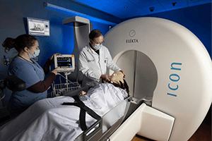 The technology itself has evolved alongside the clinical program. From the original Gamma Knife introduced in 1987 to today's Gamma Knife Esprit system, advances in magnetic resonance imaging (MRI), robotic image-guided patient positioning, patient motion management, and treatment planning have expanded the possibilities of SRS. With both frame-based and frameless treatment capabilities, UPMC continues to integrate technological innovation with decades of clinical experience, allowing increasingly individualized approaches to the treatment of complex intracranial conditions.
The effects of four decades of accumulated experience in Gamma Knife radiosurgery can be seen in how the program now approaches conditions that historically were treated differently.
One example is vestibular schwannomas. Gamma Knife can provide tumor control while preserving facial nerve function and, in selected patients, useful hearing. Experience at UPMC and through multicenter studies have subsequently shown that radiation exposure to the cochlea influences hearing outcomes. Treatment planning could then move beyond tumor control and incorporate strategies to limit cochlear dose but maintain adequate treatment of the tumor.
“At a neurosurgical meeting years ago, surgeons were shown a series of acoustic neuromas from patients who were actual neurosurgeons and asked whether they would observe, surgically remove, or use stereotactic radiosurgery as the definitive treatment option. In most of those cases, the neurosurgeons chose radiosurgery themselves as did their colleagues who were found to have these tumors as patients. That tells you how much the treatment paradigm has changed, especially when the surgical goal is to control the tumor and still preserve hearing and facial function,” Dr. Hadjipanayis says.
Brain metastases followed another similar treatment approach evolution as more long-term data became available. Whole-brain radiation therapy historically played a large role when patients developed multiple intracranial metastases, but treatment of the entire brain is known to adversely and irreparably affect memory, cognitive functions, and quality of life. Gamma Knife radiosurgery made it possible to direct treatment to individual lesions while limiting radiation exposure to uninvolved brain tissue.
“The number of metastases considered amenable to Gamma Knife radiosurgery has expanded as outcomes data accumulated,” Dr. Hadjipanayis says. “The experience progressed from patients with a single brain metastasis to those with five, 10, 20, and more lesions. Selected patients with extensive intracranial metastatic disease can now undergo stereotactic radiosurgery in scenarios in which whole-brain radiation would previously have been a more likely (and more toxic), or the only treatment pathway available. In addition, it is not uncommon for patients to undergo multiple Gamma Knife treatments for new brain metastases that are discovered with follow-up imaging of the brain.”
Arteriovenous malformations (AVMs) represent another excellent example of how the Gamma Knife has advanced treatment of these complex lesions in eloquent or functional brain areas without surgery to minimize and preserve neurologic function in patients. Larger AVMs have required another adaptation in surgical approach. Treating an extensive AVM in a single radiosurgical session can increase risk to the surrounding brain structures. The Gamma Knife program at UPMC developed staged treatment strategies in which one portion of the malformation is treated first and the remainder months later. Clinical experience has also supported repeat Gamma Knife treatment in selected patients with recurrent trigeminal neuralgia.
Meningioma management has changed as longer follow-up has become available. Small and moderate-size tumors without symptoms historically could be observed until growth or symptoms prompted treatment. Longitudinal experience has shown that selected meningiomas treated with radiosurgery can remain controlled for many years, supporting a broader role for primary Gamma Knife treatment instead of using radiosurgery only for residual or recurrent disease after surgery.
As Gamma Knife SRS became more widely adopted, UPMC also became a training site for neurosurgeons, radiation oncologists, and medical physicists learning how to use the technology in clinical practice. During Dr. Hadjipanayis’ residency, fellows and other specialists were already coming to Pittsburgh for exposure to Gamma Knife radiosurgery at a time when its role was still expanding.
Many of those clinicians later brought those experiences back to their own institutions, in many instances to different countries, where they helped establish or expand Gamma Knife programs. Over time, that created another extension of the UPMC program’s influence: the clinical experience developed in Pittsburgh informed how radiosurgery was being practiced and taught elsewhere in the world as the technology spread.
“When I trained here, Gamma Knife was still ascending as a standard treatment approach for a number of disorders, and we had people coming from around the world to learn it,” Dr. Hadjipanayis says. “Twenty years later, you can see what came from that. Many of those physicians went back to their own institutions and developed programs of their own.”
UPMC continues that work through a formal Gamma Knife training course offered six times each year. Most of the course is completed virtually, followed by two days of in-person training in Pittsburgh. Each session typically includes five or six participants from neurological surgery, radiation oncology, and medical physics who observe treatment planning and procedures with the UPMC Gamma Knife team. More than 3,000 clinicians have participated in Gamma Knife training through UPMC since the program began.
The course is structured around the same multidisciplinary approach used for patient care. Participants see how neurosurgeons, radiation oncologists, and medical physicists work through treatment planning together and how clinical, anatomical, dosimetric, and technical considerations are incorporated into the final treatment plan. That exposure is especially important for clinicians who are learning not simply how to operate the technology, but how to incorporate Gamma Knife SRS into a broader neurological and oncologic practice.
The International Radiosurgery Research Foundation (IRRF) was founded in Pittsburgh, with UPMC among its original participating institutions, and the Center for Image-Guided Neurosurgery serves as its headquarters and data coordinating center. The nonprofit consortium now includes more than 40 academic and clinical radiosurgery centers in the United States, Europe, Asia, and Africa, each represented by a team of neurosurgeons, radiation oncologists, and medical physicists. Member institutions pool outcomes data for retrospective and prospective studies across a wide range of indications. Dr. Niranjan serves as the foundation's executive director.
"We developed the International Radiosurgery Research Foundation, which brought together centers throughout the world that had Gamma Knife, to ask, 'We're seeing this in our patients. Are you seeing this too?'" Dr. Hadjipanayis says. "Now we have papers coming out almost monthly showing the long-term benefits of Gamma Knife across a number of disorders in a multicenter fashion."
Pooling data matters most for uncommon diseases and narrower clinical subgroups. Even a program that has treated more than 20,000 patients may have relatively small numbers of patients with a specific lesion, anatomical configuration, prior treatment history, or complication. Multicenter cohorts increase the number of comparable cases available for analysis, allowing questions to be answered that no single center could address on its own.
Nearly four decades after Gamma Knife arrived in Pittsburgh, its role at UPMC has become broader than that of a treatment platform. The program has provided a setting in which clinical practice, multidisciplinary care, longitudinal research, and professional training converge. The result is an experience and program that continues to change how Gamma Knife SRS is delivered, and where radiosurgery fits within the larger management of neurological disease. It also allows for questions to be asked that could not have been answered earlier in the program’s existence.
Three studies published in 2026 are illustrative of the program’s clinical maturity and longitudinal approach to data collection. These studies show exactly what kinds of questions can be answered by a program with 40 years of continuous patient care and rigorous data tracking. The three papers on long-term outcomes of Gamma Knife surgery in the setting of meningioma, dural arteriovenous fistulas, and medically refractory essential tremor examine distinct clinical problems but share a foundation: enough patients, treated and followed over enough time, to evaluate durability, safety, and changes in practice across successive eras of Gamma Knife radiosurgery.
Dr. Hadjipanayis and colleagues are now beginning to apply computational methods and artificial intelligence (AI) to the UPMC CIGNS Gamma Knife program’s prospective database to determine whether patterns accumulated over decades can help guide future treatment planning.
“We are entering a new phase with artificial intelligence. We have decades of Gamma Knife patient outcome data, and now we can begin asking whether those outcomes can help us predict the best treatment dose for a lesion based on its pathology, volume, and location,” Dr. Hadjipanayis says.
The work developed from the Computational Neurosurgery Research Group (CNRG) founded within the UPMC CIGNS by Drs. Hadjipanayis, Ajay Niranjan, and Jheremy Reyes. Building on the group's advances in AI, predictive modeling, and precision radiosurgery, Drs. Hadjipanayis, Niranjan, and Reyes established the Computational Neurosurgery Research Foundation (CNRF), an independent, international nonprofit organization dedicated to advancing computational neurosurgery through scientific research, education, technological innovation, and global collaboration. The foundation connects neurosurgeons, radiation oncologists, medical physicists, neuroradiologists, engineers, data scientists, and researchers around a shared mission: to accelerate the development, rigorous validation, and responsible clinical translation of computational technologies in neurosurgery. Through international multicenter collaborations, the CNRF supports the development and external validation of artificial intelligence models, while promoting reproducible research and evidence-based innovation.
This represents a different use of the program’s historical data. Traditional outcomes research has established how populations of patients respond to Gamma Knife radiosurgery. Computational neurosurgery takes this knowledge a step further by integrating patient-specific clinical, anatomical, imaging, and treatment data to predict individualized outcomes and explore how different treatment parameters may influence treatment response. This represents an established and rapidly expanding research effort, with the team having already published 45 studies on artificial intelligence and another 50 manuscripts currently undergoing peer review. Together, these advances are laying the foundation for a new era of precision neurosurgery, in which decades of clinical experience can be translated into increasingly personalized, data-driven treatment strategies.
The strength of the approach depends on the clinical record behind it. Artificial intelligence does not replace long-term outcome data; it depends on them. Models intended to connect lesion characteristics and treatment parameters with future outcomes require sufficiently large numbers of patients whose subsequent clinical course is known. The prospective Gamma Knife experience at UPMC provides that substrate across successive generations of imaging, treatment planning, and clinical practice.
Gamma Knife itself has also evolved across the program’s history. Imaging has become more precise, treatment planning more sophisticated, and radiosurgery has moved into clinical settings that were not envisioned when the first Pittsburgh patients were treated.
“As with any technology, Gamma Knife must keep evolving. We have been working with it for decades, so we can use what we have learned clinically to keep refining how the technology is used and how it develops,” Dr. Hadjipanayis says.
The next set of questions is therefore different from those facing Dr. Lunsford and colleagues when the program began. The initial work had to establish whether highly focused intracranial radiation could provide safe and effective disease control without the need for open surgery. Later research defined durability, adverse effects, patient selection, and the conditions for which radiosurgery could replace or complement other treatments.
Dr. Hadjipanayis and colleagues are now asking how the information generated through those decades of treatment can be used prospectively: whether previous outcomes can help predict dose, improve treatment selection, account more precisely for anatomy, and reduce treatment-related effects for the individual patient.
The more than 20,000 patients treated at UPMC represents more than a retrospective record of Gamma Knife care. Their outcomes are increasingly becoming a dataset through which the program can test how the next generation of radiosurgery should be planned.
*To read more about the process and background, interested readers can learn more in Dr. Lunsford’s recently published memoir, Brain Surgeon Without a Scalpel: Memoir of a Stereotactic Radiosurgery Pioneer, published by McFarland Press.
Learn More

