


Password Reset
Forgot your password? Enter the email address you used to create your account to initiate a password reset.


Forgot your password? Enter the email address you used to create your account to initiate a password reset.
18 Minutes
In 2021, the Division of Pediatric Nephrology at UPMC Children’s Hospital of Pittsburgh and the adult Renal-Electrolyte Division at UPMC Presbyterian were awarded a National Institutes of Health R25 grant to support a training program designed to mentor college undergraduates in kidney-based research.
Associate professor of Pediatrics Sunder Sims-Lucas, PhD, who studies the basic science of acute kidney injury (AKI), leads the program, along with clinician-scientist and Division chief Jacqueline Ho, MD, MS, who specializes in the study of acute kidney injury, kidney disease, and how microRNAs influence disease mechanisms and processes.
The R25 award supports an innovative undergraduate training program called “Summer Research Internship Program Kidney Workshop (SRIP-Kid).”
The training program, having recently completed its second full year, provides undergraduate students interested in kidney-based research to travel to the University of Pittsburgh and take part in a 8-week program where they conduct research and receive didactic instruction on a range of topics in nephrology from researchers and physicians in the Division of Pediatric Nephrology and collaborators in the adult Renal-Electrolyte Division and Department of Critical Care Medicine at the University of Pittsburgh School of Medicine.

“Our first couple of years of the program were disrupted by the COVID-19 pandemic, but this year we were able to have a much more robust and in-person experience for our students,” says Dr. Sims-Lucas. 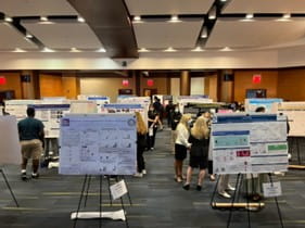
While the program affords participants a unique opportunity to gain hands-on laboratory experience and be mentored by leading nephrology researchers working in various laboratories at UPMC and the University of Pittsburgh, a novel aspect of the program is that students have the opportunity to take part in clinical activities and experiences that match up with the type of research in which they are engaged.
"For example, one of our students is an actual kidney transplant recipient who was able to take part in a transplant-related research project, but also observe and take part in transplant clinics and rounding sessions at the hospital," says Dr. Sims-Lucas. "The kind of experience that our program provides shows these students that they can have careers working in the basic or translational laboratories, but also the path of being a clinician-scientist, treating patients but also engaged in biomedical research."
Didactic education developed for the program includes a kidney-specific workshop where students learn the fundamental aspects of kidney formation, physiology and cellular functional mechanisms, and various kidney diseases – how and why they occur and how they operate on molecular and cellular levels to cause renal dysfunction.
At the end of the course, students present their research in a scientific poster session [see below for abstracts] to other students in the program and faculty members from various Divisions and Departments at the University of Pittsburgh School of Medicine and UPMC. Also, part of their training is a workshop led by pediatric pulmonary medicine researchers Michelle Manni, PhD, that teaches students how to write a research abstract and give a poster presentation.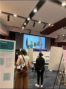
“Additionally, four of our students in the kidney program are selected to travel to the American Society of Nephrology’s annual Kidney Week conference with me to be immersed in all of the cutting-edge basic science, translational, and clinical work that will be showcased by nephrology colleagues from all around the world," says Dr. Sims-Lucas. "It's a fitting coda to the work these students performed in our program to see what happens globally in terms of kidney research."
The SRIP-Kid program is evolving into a robust early training ground for young students who may one day go on to help solve some of the most complex kidney disease challenges facing clinicians and patient alike. The magnitude and scope of the program are truly a collaborative and team effort.
“I’d like to give a special thanks to everyone in our pediatric and adult nephrology divisions at the University of Pittsburgh and UPMC who give their time and knowledge in mentoring our students,” says Dr. Sims-Lucas. “The program also is indebted to the work of the team running the University of Pittsburgh Summer Research Internship Program, including Nama Ballas, as well as Diane Klein and Margaret Jaczesko.”
Further Reading – Research Abstracts
Readers interested in the scientific abstracts from the summer research projects can find them in their entirety below. Names in yellow are the student authors.
3D Modeling of Dense Apical Tubules in Renal Proximal Tubules
Authors: Isabella Cowan; Dr. Ora Weisz; Renal-Electrolyte Division, University of Pittsburgh School of Medicine
The apical endocytic pathway of kidney proximal tubule (PT) cells has a unique organization that is presumably needed to maintain the high endocytic capacity of this nephron segment. Megalin and cubilin receptors on these cells bind to proteins and other ligands that escape the glomerular filtration barrier and mediate their uptake into endocytic compartments. Bound ligands are dissociated from receptors in these apical endosomes and delivered to lysosomes for degradation, while the receptors are collected in membrane-rich dense apical tubules (DATs) that bud from endocytic compartments for recycling to the apical surface. How the organization and morphology of apical endocytic compartments contributes to the high endocytic capacity of PT cells is not known. We sought to examine this question using a recently acquired Focused Ion Beam Scanning Electron Microscopy (FIB-SEM) dataset that features high-resolution scans of mouse renal PT cells. Specifically, we sought to determine whether DATs form an interconnected network as opposed to unique structures. Additionally, we hoped to identify the site of DAT fusion with the apical plasma membrane. We used Zeiss APEER software to manually annotate DAT structures in a subset of FIB-SEM sections, then used deep learning (DL) segmentation to automatically identify all DATs in the training set. This created a prediction model that we then imported into Arivis Vision4D, allowing us to confirm accuracy and fine tune the model through analysis pipelines (filtering by pixel size, color, or proximity to other pixels, denoising, and further segmentation) to generate a final 3D representation of the arrangement of DATs within the PT. Progress on this ongoing research will be presented.
Persistent DNA Damage Causes Renal Metabolic Reprogramming and Leads to Chronic Kidney Disease
Authors: Haley Arbore1, Amy B Huynh2, Merlin Airik2, Rannar Airik2,3. 1Department of Biological Sciences, University of Pittsburgh, PA; 2Division of Nephrology, Department of Pediatrics, University of Pittsburgh, PA; and 3 Department of Developmental Biology, University of Pittsburgh, PA
Chronic kidney disease (CKD) is a significant health concern affecting >10% of global population. In the US, CKD affects >37 million people and costs > 30 billion per year in health care dollars. Despite its high prevalence, we have few therapies to slow or halt the progression of kidney damage in CKD. Thus, understanding mechanisms that drive CKD progression is critical for impacting patients. Emerging evidence suggests that impaired DNA repair and accumulation of DNA damage in the kidney proximal tubular cells (PTECs) are involved in CKD. However, the mechanism by which accumulation of DNA damage leads to tubular injury is unclear. To address this, our lab generated a DNA repair deficient Fan1KO mouse model which is orthologous to the human hereditary kidney disease, karyomegalic interstitial nephritis (KIN).
Using the Fan1KO mouse model, I show that persistent DNA damage in the kidney tubular epithelium after cisplatin injury leads to metabolic reprogramming in the tubular cells, which is characterized by reduced fatty acid oxidation and increased aerobic glycolysis. Glycolytic shift in Fan1KO kidneys was associated with increased levels of reactive oxygen species (ROS), damage to mitochondria and de novo expression of glycolytic enzymes in the proximal tubules, as demonstrated by immunofluorescence and immunohistochemistry analyses of the kidneys. Western blotting of glycolytic enzymes and their upstream regulators Akt, mTOR and AMPK further demonstrated that persistent DNA damage leads to metabolic reprogramming in tubular cells of the kidney. Histological analysis showed progressive tubular injury in Fan1KO kidneys after cisplatin injury, consistent with the development of chronic kidney disease. Treatment with a mitochondrial ROS scavenger attenuated the glycolytic shift and mitigated CKD pathogenesis in Fan1KO kidneys, suggesting that mitochondrial deficiency is an integral component in DNA damage driven CKD. Together, my data suggest that accumulation of DNA damage in the kidney proximal tubule cells causes tubular metabolic reprogramming which leads to tubular injury and development of chronic kidneys disease. Future work will investigate the molecular mechanisms by which tubular DNA damage affects mitochondrial energy metabolism in order to prevent or mitigate mitochondrial damage in CKD patients and prevent further kidney damage.
Entrance Skin Dose Characterization for the Stochastic Risk of Ionizing Radiation Exposure in Abdominal X-ray Imaging of Pediatric Patients
Authors: Alyssa Paul1; Rajeev Chaudhry, MD2. 1Intern: Department of Urology, Butler University, UPMC Children’s Hospital of Pittsburgh; 2Advisor: Department of Urology, University of Pittsburgh School of Medicine, UPMC Children’s Hospital of Pittsburgh
Background: X-ray imaging is a popular diagnostic tool utilized by pediatric doctors. X-rays expose patients to ionizing radiation, which children have a greater sensitivity to due to their rapid cell division, constant growth, and long-life expectancy. Currently, dose area product (DAP) is used to estimate skin level radiation exposure. DAP is not considered accurate by the United Nations Scientific Committee on the Effects of Atomic Radiation (UNSCEAR) since it is calculated from air kerma rather than true entrance skin dose. DAP also assumes all patients have the same sex, age, and body mass index.
Objective: The objective of this study was to measure the entrance skin dose for abdominal X-rays on pediatric patients to assess the accuracy of air kerma readings.
Study Design: The study was done prospectively as 35 pediatric urology patients underwent abdominal X-ray imaging. Entrance skin dose was measured using Landauer NanoDot™OSLD dosimeters adhered to the navel with hypoallergenic tape. X-ray technicians recorded the presets for each study, including air kerma values. A null hypothesis of no statistically significant difference between the air kerma and measured skin level radiation values was used when testing data. Statistical analysis was performed with Excel and MATLAB.
Results: Patients had a median age of 9 years (IQR 5.5-13) and a median BMI in the 52.35th percentile (IQR 35.54-94.135). The median air kerma and entrance skin dose were 0.562 mGy (IQR 0.219-1.13) and 0.752 mGy (IQR 0.299-1.72) respectively. A paired, two-tailed t-test found air kerma and entrance skin dose to be significantly different (p=0.0009). There was a positive correlation between air kerma and error, with a Pearson coefficient of 0.745. Chi square analysis found entrance skin dose is dependent on both age and BMI (p=2.37E-05 and p=8.14E-06, respectively). There is a positive correlation between age and entrance skin dose, with a Pearson coefficient of 0.480. BMI and entrance skin dose also have a positive correlation, with a Pearson coefficient of 0.350.
Conclusions: Abdominal X-rays appear to be a low-risk procedure with minimal radiation exposure for diagnostic medicine. Measured radiation absorption at skin level is greater than the air kerma value from which effective dose is calculated. With increase in age and size, higher radiation doses are typically needed for penetration to produce effective results. This is done by increasing voltage, which increases the radiation dosage. As air kerma increased, inaccuracy of air kerma increased. This suggests absorbed radiation is greater than currently estimated in air kerma values when multiple X-rays at higher intensities are taken. Taking less images or using lower dose settings can help to limit the stochastic risk of pediatric patients further.
Impact of HIF-1α on Nrf2 Activity in a HK-2 Cell Culture Model of Acute Kidney Injury
Authors: Joy A. Stewart, Corry D. Bondi, PhD, MS, and Roderick J. Tan, MD, PhD
University of Pittsburgh, Department of Medicine, Renal-Electrolyte Division
Acute kidney injury (AKI) is a rapid decline in kidney function that can occur after ischemia/reperfusion injury (IRI) and may lead to chronic kidney disease or end-stage kidney disease. Currently, there is no effective treatment for IRI-AKI, thus it is crucial to establish a better understanding of its pathogenesis. To mitigate the effects of ischemic conditions, cells have evolved adaptive cytoprotective responses such as the nuclear factor erythroid-2-related factor 2 (Nrf2) and hypoxia-inducible factor-1α (HIF-1α). Nrf2 activates protective detoxifying and antioxidant genes in response to cellular stress. Several studies show that Nrf2 activation protects against ischemic AKI. However, our lab showed a maladaptive decrease in Nrf2 activity in severe AKI in mice. The mechanism behind this is unknown. Interestingly, HIF-1α is upregulated in IRI-AKI and may influence Nrf2 activity. I hypothesized that HIF-1α inhibits Nrf2 activity through an intervening signaling pathway. To investigate my hypothesis, I utilized an in vitro cell culture system to simulate severe ischemic conditions. Because ischemia impairs nutrient and oxygen delivery, I exposed immortalized proximal tubule epithelial cells (HK-2 cells) to nutrient deficient (Hanks balanced salt solution) conditions in the presence or absence of the HIF-1α activator, CoCl2. CoCl2-mediated HIF-1α activation inhibited Nrf2 activity, as evidenced by a reduction in NQO1 mRNA expression. I interrogated potential pathways linking HIF-1α to Nrf2 by using CX4945, LY294002, and U0126, to inhibit casein kinase 2, MEK/ERK 1/2, and PI3K/AKT pathways, respectively. No significant differences in NQO1 mRNA expression were found when these pathways were inhibited. This suggests that these pathways were unable to block the suppressive effect of HIF-1α on Nrf2 and most likely are not involved. Future studies should continue to explore other kinase pathways such as p38, JNK, PKC, and GSK3-β, and proteins involved in the nuclear trafficking of Nrf2.
The Effects of FRMD6 Overexpression on the TGF-β and Hippo Signaling Pathways
Maliha Tayeb1; Débora Malta Cerqueira1; Andrew Bodnar1, and Jacqueline Ho1
1. Division of Nephrology, Department of Pediatrics, UPMC Children’s Hospital John G. Rangos Sr. Research Center, Pittsburgh, PA
Chronic Kidney Disease (CKD) is an increasingly prevalent disease that affects approximately 37 million people in the United States. It is characterized by the long-term development of renal fibrosis: the excessive deposition of extracellular matrix (ECM) proteins, such as collagen. The transforming growth factor (TGF-β) and Hippo signaling pathways can lead to increased ECM protein production through the activation of downstream Smad proteins that enhance the transcription of pro-fibrotic target genes. Our laboratory studies microRNAs, which are small, non-coding RNAs that inhibit post-transcriptional gene expression by binding to specific mRNA targets. We have previously demonstrated that the inducible deletion of the microRNA cluster miR-17~92 in proximal tubules, distal tubules, and collecting ducts results in increased renal fibrosis, following unilateral ureteral obstruction in a mouse model. We have preliminary data that demonstrates that overexpression of FRMD6 (a member of the Hippo signaling pathway and proposed miR-17 target) in HK-2 cells induces increased collagen secretion into conditioned media. In this study, we tested the hypothesis that FRMD6 is a miR-17 target and FRMD6 overexpression results in activation of the pro-fibrotic TGF-β and Hippo signaling pathways. We used two in vitro models in HK-2 cells that we predict will result in FRMD6 overexpression: transfection with pCMV_Sport6 vector containing FRMD6 cDNA and transfection with miR-17 inhibitor. Immunostaining confirmed the overexpression of FRMD6 using pCMV_Sport6 vector with FRMD6 cDNA. To assess the downstream effects of FRMD6 overexpression within the TGF-β and Hippo signaling pathways, we harvested HK-2 cells transfected with FRMD6 cDNA via nucleus and cytosol isolation to evaluate the levels of phosphorylated Yes-Associated protein (p-YAP) in the Hippo signaling pathway that may interact with phosphorylated-Smad (p-SMAD) protein in the TGF-β signaling pathway. Although we predicted increased p-YAP in cells transfected with FRMD6 cDNA, the western blot of this protein within the nucleus and cytosol did not demonstrate a significant difference. Overall, further experiments are needed to determine if miR-17 targets FRMD6 and if there is a role for FRMD6 in renal fibrosis. Future directions include carefully defining tissue culture conditions for transfection of HK-2 cells, evaluating the efficiency of the miR-17 inhibitor, and assessing if overexpression of FRMD6 causes other detrimental effects, such as apoptosis, that may confound our interpretation of the results.
Influence of MUC1 upon Apical Localization of Ca2+-Selective TRP Channels In-vivo
Authors: Lorena Ye, Tracey Lam, Allison Marciszyn, Mohammad Al-Bataineh, Becky Hughey, Evan Ray, Department of Medicine, University of Pittsburgh
Background: MUC1 is a heavily glycosylated single transmembrane protein expressed on the apical surface of most mammalian epithelia. In the kidney, MUC1 is expressed in the distal nephron from the thick ascending limb of the loop of Henle into the collecting duct. It is also expressed throughout the GI tract. Previous studies in cell culture show that MUC1 increases the expression and activity of Ca2+-selective channel TRPV5. We find that mice lacking MUC1 have lower blood calcium levels.
Hypothesis: We hypothesize that MUC1 knockout mice will have reduced apical localization of TRPV5 and TRPV6 in the kidney and intestine.
Methods: Male and female MUC1+/+ and MUC1-/- mice were sacrificed at 16 to 20 weeks of age, and kidneys and duodena were removed at time of sacrifice. 5 μm-thick cryosections were prepared and immunostained. Immunolabeled tissues were imaged using a confocal laser scanning microscope (Leica TCS SP5, Model upright DM 6000S, Leica Microsystems Inc., Buffalo Grove, IL, USA), and the resulting images were analyzed in Image J using line scans to measure TRP channel staining intensity across the apical membrane. Line scan intensity profiles were normalized to peak intensity using IgorPro software, and profiles were compared between MUC1+/+ and MUC1-/- mice. Cytoplasmic/apical staining intensities were calculated and compared between genotypes using Student’s T-test.
Results: In the distal convoluted tubule, MUC1-/- mice exhibited redistribution of TRPV5 and TRPV6 from the cell apex to the cytoplasmic space. In the duodenum, TRPV6 was similarly redistributed toward the cytoplasm.
Conclusions: Reduced apical/cytoplasmic localization of Ca2+-selective TRP channels provides a mechanism to explain reduced blood Ca2+ in MUC1-/- animals. This may occur through reduced Ca2+ absorption in the GI tract or through reduced Ca2+ reabsorption in the distal convoluted tubule.
BK Channel Alpha Splicing Variants in Mouse Kidneys During Development and Dietary K+ Challenge
Authors: Agnes Han, Stephanie Mutchler, Catherine Priestley, Shujie Shi and Thomas Kleyman
Renal-Electrolyte Division, Department of Medicine, University of Pittsburgh, Pittsburgh, Pennsylvania
BK channels are calcium or voltage activated K+ channels that mediate K+ secretion and homeostasis in the distal nephron and are highly expressed in intercalated cells (ICs). BK channels are tetramers of an α pore-forming subunit, encoded by the KCNMA1, or SLo1 gene, which contains 35 exons. BK channels achieve phenotypic diversity despite only being encoded by a single gene through substantial alternative splicing of the α subunit. How αBK splice variant expression in the kidney and ICs is affected by increases in dietary K+ or by changes during development is still unknown. The current study investigates changes in BK variant expression during development and under a high K+ diet at two sites of interest: the cytosolic carboxyl-terminus (C-terminus), and another complex splice site between exons 20 and 24. It further evaluates differences in variant abundance between the whole kidney and ICs at seven identified splice sites. Adult C56B6 mice were fed with the Ctrl or a 10% KCL diet for 9 days and sacrificed at 8-12 weeks. Total RNA was isolated from postnatal-day (PD) 0, 7, 14, adult high K+ and adult control mice kidneys. Quantitative RT-PCR was used to determine expression levels for each splice variant. IC isolation was carried out by crossing V-ATPase B1-cre mice to tdTomato-loxp reporter mice to drive tdTomato expression in B1-positive renal ICs. IC population was then enriched by fluorescence-activated cell sorting and used for total RNA isolation, followed by reverse-transcriptase (RT) PCR and gel electrophoresis. For mice placed on a high K+ diet compared to control mice, expression of variants ERL, VYR and 22 increase, whereas expression of variant zero remains constant. For mice at different developmental stages, disparate effects were observed for male versus female mice. For all splice variants at both sites under investigation, female mice upregulate BK expression over time from P0 to adulthood. Male mice likewise steadily upregulate expression of variants zero from P0 to adulthood. However, for variants VYR, ERL, and e22, male mice demonstrate similar levels of expression at P0 and adulthood, with upregulation from P0 to P14 followed by downregulation from P14 to adulthood. Knowledge gaps remain: both the differences in variant expression observed between male and female developing mice and the changes in variant expression specifically in ICs when challenged with dietary K+ intake warrant further investigation.
Dicarboxylic Acid Enriched Diet Protects Against Renal Fibrosis in a CKD model.
Authors: Adham Abdo and Sunder Sims-Lucas. Department of Nephrology
Chronic Kidney Disease (CKD) is characterized by fibrosis of the renal interstitium decreasing kidney functionality. Currently, a diagnosis of CKD usually occurs only after the fibrosis has become severe enough to cause significant cell death, limiting the plausible clinical treatments to transplant or life-long dialysis. We investigated a potential therapeutic strategy to halt or regress the progression of renal fibrosis, which could vastly improve the lives of people living with, or at risk for CKD. We have previously seen that a diet enriched in medium-chain fatty acids (10% dodecanedioic acid) has been protective in Acute Kidney Injury. We hypothesize this protection is due to a shift in the metabolism in the proximal tubule epithelial cells. The addition of medium chain dicarboxylic acid (DCA) increases the peroxisomal-specific FAO substrates, limiting the damaging effects of Reactive Oxygen Species-mediated oxidative stress that can result from mitochondrial FAO. Here we show a similar protection in a CKD model. Mice pretreated with a DCA-enriched diet or control diet underwent a Unilateral Ureter Obstruction (UUO). Through histological stains and immunofluorescence, we show a decrease in fibrosis markers in those mice pretreated with DCA. We suggest that increased peroxisomal activity through supplementation with medium-chain dicarboxylic acid is a potential clinical treatment for CKD.
