

Password Reset
Forgot your password? Enter the email address you used to create your account to initiate a password reset.


Forgot your password? Enter the email address you used to create your account to initiate a password reset.
6 Minutes
The Ferguson Laboratory for Orthopaedic and Spine Research has been using human derived cells and tissues for years to study the basic biology of spine degeneration and chronic low back pain, among other lines of investigation. Access to the growing Bethel Musculoskeletal Research Center (BMRC) Biobank is now allowing the laboratory to expand its research options utilizing multiple human musculoskeletal tissues for tissue explants, organoids, bioreactors, and other experimental model systems.
These expanded options for using human tissues in research coincides with greater emphasis from the National Institutes of Health on incorporating human-derived models into biomedical research. Within the Ferguson Laboratory, the shift provides flexibility as well as an opportunity to develop more human-relevant models while continuing to use animal models for questions that require an intact biological system or which simply cannot be done in human tissues or in human clinical studies.
Animal models have long allowed scientists to investigate disease mechanisms and evaluate potential interventions before they can be studied in people. However, a biological pathway or finding identified in an animal model may resemble but not precisely reproduce the corresponding pathway in humans. Those differences can contribute to the failure of drugs and other interventions when they move from preclinical studies into human trials.
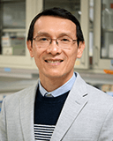 “Animal models can bring us closer to understanding a disease process, but they may not bring us close enough,” says Nam Vo, PhD, professor of orthopaedic surgery and co-director of the Ferguson Laboratory. “A study can identify something important in an animal and still lack additional information needed to understand what will happen in a person.”
Biological variation among people introduces further complexity. Patients with the same diagnosis may differ in their genetics, tissue biology, environmental exposures, and other sociopsycho characteristics that influence disease progression and treatment response. Human-derived models give scientists another way to investigate some of these differences using cells and tissues obtained directly from patients.
The greater emphasis on human-derived models should not be interpreted as evidence that animal studies are no longer needed. Human cell cultures, explants, organoids, and bioreactors each reproduce selected elements of human biology, but none recreates an entire person. Questions involving systemic physiology, immune responses, metabolism, or interactions among multiple organs may still require an animal model.
“The goal should be to determine which model is appropriate for each scientific question,” Dr. Vo says. “Many projects will benefit from combining several models so that the strengths of one compensate for the limitations of another.”
The Ferguson Laboratory has used intervertebral disc tissue collected during surgery for a number of years. Lab staff isolate cells from the tissue, expand them in culture, and use them to examine cellular processes associated with spine degeneration. The laboratory also derives cells from ligamentum flavum surgical specimens.
The BMRC Biobank provides a more extensive and systematic source of human musculoskeletal tissue. Established procedures govern patient consent, collection in the operating room, transport, processing, preservation, specimen tracking, and research access. The proximity of UPMC operating rooms, the Biobank, and Pitt research laboratories also allows specimens to be transferred and processed promptly, including when studies require fresh tissues or viable cells.
That infrastructure addresses an obstacle faced by scientists at institutions where surgical services and research laboratories are separated by considerable distance or where no coordinated tissue collection process exists. It also gives the Ferguson Laboratory access to several tissue types needed to construct models that extend beyond an isolated population of, for example, intervertebral disc cells.
The Ferguson Laboratory is working toward organoid and bioreactor approaches that incorporate human cells derived from several components of the spine. These include the intervertebral disc, paraspinal muscle, vertebral bone, and ligamentum flavum.
“We are trying to design a chip that allows us to grow cells from those different tissues together and study how they interact,” Dr. Vo says. “These are all human cells. Without the BMRC Biobank and access to the different orthopaedic tissues, developing that type of model would be very difficult.”
A culture containing disc cells alone can reveal how those cells respond to a defined stimulus. It cannot show whether signals originating in muscle, bone, or ligament tissue alter that response. The model would still reproduce only selected features of the spine. It would not contain the complete vascular, neurological, immune, and mechanical environment found in a person. Instead, it would provide an important intermediate model between isolated cell culture and whole-organism research, allowing the laboratory to study interactions that single-tissue models cannot capture.
The BMRC Biobank also permits several forms of analysis beyond cell culture. Fresh tissue can provide viable cells, while frozen specimens can support subsequent molecular studies. Paraffin-embedded tissue can be used for histology and other imaging-based analyses. Genomic, epigenomic, proteomic, and other multiomic methods can add further information about the biological characteristics of each specimen.
Those findings can then be evaluated alongside data from the patient. Relevant information may include MRI examinations, radiographs, clinical history, treatments, and longitudinal outcomes documented in the electronic health record.
“No individual dataset provides the entire answer,” Dr. Vo says. “Imaging shows structural characteristics. The electronic health record provides clinical history, diagnoses, treatments, and outcomes. Tissue analyses reveal cellular and molecular features.”
Integrating these sources may help the Ferguson Laboratory investigate why patients with apparently similar clinical presentations follow different courses. Two patients with chronic low back pain may have different underlying biological processes, rates of progression, or responses to treatment. As these datasets grow, machine-learning methods may help identify combinations of biological, imaging, and clinical characteristics associated with disease trajectory or recovery.
Greater use of human specimens also raises a related question: how can institutions use animal models more efficiently when they are necessary for specific research projects or needs?
A laboratory studying one organ or tissue may collect that material and discard the rest. That is inefficient at best and wasteful at its worst. This is particularly consequential with aged animals, which require substantial time and expense to maintain. Dr. Vo previously led an aging working group at Pitt that coordinated tissue collection among laboratories. When aged models were used, participating laboratories collected different tissues from the same animals, making the most efficient use of an expensive research asset.
“It is not only a matter of reducing the use of animals,” Dr. Vo says. “We also need to use them more effectively in a collaborative and integrated manner. It becomes a matter of conducting the best science possible while optimally managing the resources at your disposal. It’s about effective stewardship, ethical principles, and the responsible use of using living organisms for scientific discovery.”

